Image compression for teleradiology: a buyer's guide
Image compression for teleradiology explained for buyers: lossless vs lossy, ACR guidance, FDA limits, and what to put in your contract and SLA.
Image compression for teleradiology is the process of shrinking DICOM studies so they travel faster to a remote radiologist and take less room in storage. Reversible (lossless) compression leaves every pixel unchanged and is the safe default. Irreversible (lossy) compression is acceptable only when image quality stays sufficient for the specific clinical task, and that call belongs to a responsible physician, not a vendor's default setting.
TL;DR
- Lossless compression typically shrinks studies by 1.5:1 to 3.6:1, while lossy methods reach 5:1 to 50:1 at the cost of discarded data, according to a 2020 RANZCR guideline.
- The 2022 ACR,AAPM,SIIM technical standard says irreversible compression is acceptable only when image quality is sufficient for the clinical task, with physician-directed settings.
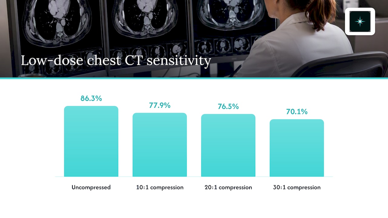
- A 2003 Radiology study of low-dose chest CT found sensitivity fell from 86.3% uncompressed to 70.1% at 30:1 JPEG 2000 compression.
- The FDA does not allow irreversible compression of digital mammograms for retention, transmission or final interpretation.
- AstraRad delivers final signed reports straight into your existing PACS, DICOM conformant, with pricing per report and no minimums.
Table of Contents
- What is image compression in teleradiology, and why does it matter?
- Who are we, and what is our interest in this topic?
- How does image compression for teleradiology work?
- What is the difference between lossless and lossy compression?
- Can lossy compression change a diagnosis?
- What do US rules say about compression for diagnostic reads?
- What should you check when transmitting compressed images to a remote reader?
- Which compression approach fits which setting?
- How does AstraRad fit into your compression and PACS workflow?
- What should a teleradiology contract say about compression?
- How do you validate compressed images before go-live?
- Is compression still necessary with modern networks?
- Where does AstraRad's offer fit?
- FAQ
- Sources
What is image compression in teleradiology, and why does it matter?
Image compression is a technique that reduces the number of bytes needed to store or send a digital image. In teleradiology it matters because every study has to cross a network before a radiologist can read it, and file size drives both transmission time and storage cost.
The size problem has grown, not shrunk. A 2021 review of medical imaging compression standards notes that a modern thoracic CT can run 600 MB to 1 GB, compared with roughly 12 MB for a typical thoracic CT in the early 1990s. Multiply that by an overnight queue and the bandwidth question becomes a procurement question.
Compression is also old news in the literature. A 1995 telemedicine study reported JPEG files at about 10% of original size with no noticeable quality change in its tested X-ray, CT and ultrasound samples, which implies roughly a 90% cut in transmission time. That result is a reason to take compression seriously, not a license to apply it everywhere, as the next sections show.
Three terms come up in every vendor conversation:
- DICOM is the standard format and network protocol for medical images and their metadata.
- PACS is the picture archiving and communication system where your studies are stored, viewed and reported.
- Compression ratio is the original file size divided by the compressed size, so 10:1 means the file is one tenth its original size.
Who are we, and what is our interest in this topic?
AstraRad is a US teleradiology service that delivers final signed reports read by board-certified subspecialists. We sell per-report teleradiology to hospitals, imaging centers and radiology groups, so we have a commercial interest in you choosing a vendor like us, and you should read this guide with that in mind.
We wrote it because compression settings sit between your PACS and any remote reader, and they rarely appear in a contract. Our reports go straight into your existing PACS with no extra portal, and our workflow is DICOM conformant. You can see the full path from your PACS to a signed report on our how teleradiology works page, and the range of modalities we read on our teleradiology services page.
How does image compression for teleradiology work?
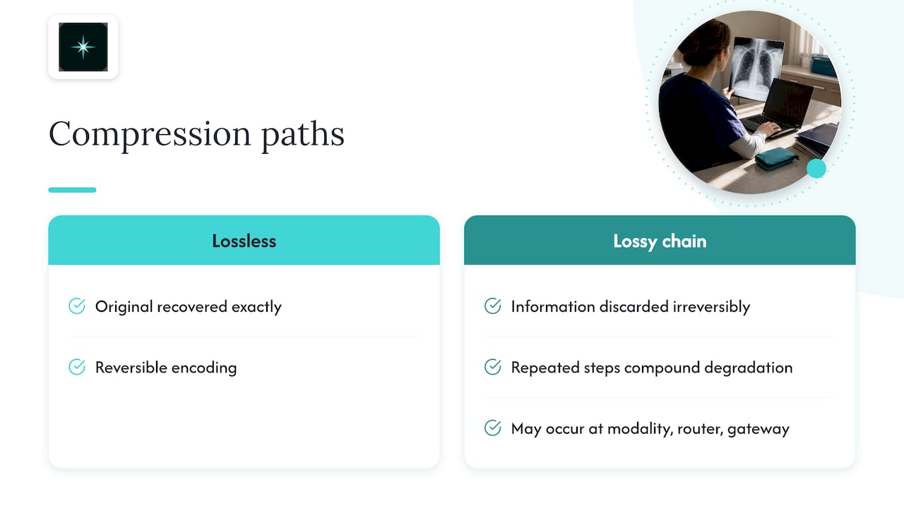
Compression works by encoding the pixel data of a DICOM image more efficiently. Lossless methods find redundancy and remove it in a way that can be fully reversed. Lossy methods also discard information the algorithm judges least important, so the original can never be recovered exactly.
DICOM defines specific algorithms for this. The ACR,AAPM,SIIM standard recommends DICOM-defined algorithms such as JPEG, JPEG-LS, JPEG 2000 and MPEG. It warns that proprietary schemes impede interoperability, and that repeated decompression and irreversible recompression can significantly degrade quality.
That last point matters in a teleradiology chain. A study may be compressed at the modality, again at a router, and again by a cloud gateway. Each lossy step compounds the one before it. A buyer who asks only for the "compression ratio" misses the real question: how many times, by which method, and where in the chain?
What is the difference between lossless and lossy compression?
Lossless compression is a reversible method: decompression reproduces the original image exactly. Lossy compression is an irreversible method that removes data and may introduce distortion. The tradeoff is file size against fidelity, and the numbers below come from the RANZCR guideline.
| Feature | Lossless (reversible) | Lossy (irreversible) |
|---|---|---|
| Original image recoverable | Yes, exactly | No |
| Typical ratio | 1.5:1 to 3.6:1 | 5:1 to 50:1 |
| Image quality impact | None | Depends on ratio, modality, body part and task |
| Main risk | Smaller bandwidth savings | Artifacts that hide or mimic findings |
| Typical use in remote reads | Default for primary interpretation | Limited, task-specific, physician-approved |
The European Society of Radiology's review adds a useful distinction. Lossy JPEG and JPEG 2000 may be visually lossless or diagnostically lossless depending on modality, anatomy, disease and task. It stresses that visually lossless does not guarantee diagnostically lossless, especially for low-contrast detection, fine texture and artifact-prone findings.
In plain terms, an image can look identical to a reader on a quick comparison and still have lost the subtle finding that matters. That is why the standards focus on the task, not on the ratio.
Can lossy compression change a diagnosis?
Yes, at high enough ratios and for some tasks. Studies show lossy compression can be harmless at modest ratios for some images and measurably harmful at higher ratios for others. The result depends on the modality, the finding and the algorithm, so no single ratio is safe everywhere.
Three published studies show the range:
- Body CT, 2000. A study of JPEG and wavelet compression of body CT found 10:1 JPEG compression suitable for on-call transmission, provided the original images were subsequently reviewed. At 20:1 some tested measures suffered.
- Low-dose chest CT, 2003. A Radiology study of wavelet compression reported sensitivity of 86.3% uncompressed, 77.9% at 10:1, 76.5% at 20:1 and 70.1% at 30:1, even though specificity stayed above 98%. Lung nodule detection is a low-contrast task, and it showed.
- Mammography, 2015. A 2015 AJR study found no statistically significant difference in reader performance between JPEG 2000 at 80:1 and non-lossy images. It also noted that federal regulations prohibited lossy compression for primary reading and archiving.
Read together, these results say two things. First, "it worked in one study" is not evidence for your protocol. Second, a clinical-grade program treats any lossy step as a documented clinical decision, with the original available for review when a finding is in doubt.
What do US rules say about compression for diagnostic reads?
In the US, the main professional guidance is the ACR,AAPM,SIIM technical standard for electronic practice of medical imaging, updated in 2022. It says reversible compression has no image-quality impact and that irreversible compression is acceptable only when image quality remains sufficient for the specific clinical task.
The same standard defines diagnostically acceptable irreversible compression as compression that does not reduce diagnostic performance for the primary interpreter or other decision makers, with artifacts imperceptible or non-interfering. It makes no universal compression-ratio recommendation. The acceptable amount depends on body part, modality and the study's objective.
Who decides? The standard places initial selection and periodic review of compression settings with the responsible physician, working with a qualified medical physicist as appropriate. For a buyer, that means your medical director or radiology lead owns the policy. A vendor can implement it, but should not set it silently.
What about mammography?
Mammography is the clearest hard line. The ACR standard states that the FDA does not allow irreversible compression of digital mammograms for retention, transmission or final interpretation. FDA-cleared PACS documentation says the same thing in practice: lossy-compressed mammographic images must not be used for primary diagnostic interpretation, and mammography requires approved digital-mammography displays.
If you outsource screening or diagnostic mammograms, confirm that the whole chain, from modality to remote workstation, stays lossless. Our telemammography page describes how we handle MQSA-qualified remote reads.
What should you check when transmitting compressed images to a remote reader?
Check five things: which algorithm is used, how many times images are recompressed, whether bandwidth matches your volume, whether compression is labeled on the image, and whether the receiving system is DICOM compatible. These come directly from the ACR,AAPM,SIIM standard and the 2021 standards review.
- Use DICOM-defined algorithms. Avoid proprietary schemes. The 2021 review states that DICOM interoperability and compatibility should drive the choice of compression standard, rather than compression efficiency alone.
- Avoid repeated irreversible recompression. Map every hop between modality and reader and confirm only one place, if any, applies a lossy step.
- Match bitrate and bandwidth to expected volumes. Size the link for your peak overnight and backlog scenarios, not your average day.
- Label compression on the image. The standard states that displayed images should identify irreversible compression, give an approximate ratio or quality factor, and name the scheme. That information should persist after decompression.
- Use relevant IHE profiles. They give both sides a shared, testable way to exchange studies.
A practical test is simple. Ask for a sample study sent end to end, then compare the received DICOM header against the original. If the transfer syntax differs from what you expected, you have found a conversation worth having before go-live.
Which compression approach fits which setting?
The right approach depends on the modality, the clinical task and your network. The table below summarizes how the standards and studies cited in this guide translate into buyer decisions. It is a starting framework for your responsible physician, not a substitute for their judgment.
| Situation | Safer approach | Why |
|---|---|---|
| Digital mammography | Lossless only | The FDA does not allow irreversible compression for retention, transmission or final interpretation |
| Primary diagnostic read, any modality | Lossless by default | Reversible compression has no image-quality impact, per the ACR,AAPM,SIIM standard |
| Low-contrast tasks such as lung nodule detection | Avoid lossy | Sensitivity dropped at 10:1 and beyond in the 2003 chest CT study |
| Emergency preview over a weak link | Lossy only if physician-approved, with originals reviewed afterward | The 2000 body CT study supported on-call use at 10:1 on that condition |
| Long-term archive | Lossless, unless a physician-approved policy says otherwise | Archived images must remain diagnostic for later comparison |
Notice that the ratios in these studies are not rules for your site. They show how fast performance can change, which is why the ACR standard asks for periodic review of settings instead of a one-time choice.
How does AstraRad fit into your compression and PACS workflow?
AstraRad is a US teleradiology service that delivers final signed reports read by board-certified subspecialists. Each study is routed to a subspecialist matched to the modality and body part, and every read is performed inside the United States on scheduled shifts, 24/7/365.
For your IT team, the relevant points are integration and accountability. Reports return directly to your existing PACS with no extra portal, and the workflow is DICOM conformant. We work as a business associate under a signed BAA, and we are HIPAA aligned. Turnaround is committed by contract and measured from last-image arrival to radiologist signature: STAT under 4 hours (under 2 hours or under 1 hour on request), routine 24 to 48 hours, and stroke protocol under 30 minutes.
Pricing is per report with no minimums. You can request a full per-report rate card at /pricing, or book a discovery call at /book-a-call. A coverage plan with SLA terms in writing follows the call, which is the right place to document your compression policy.
What should a teleradiology contract say about compression?
A teleradiology contract should state which compression types are permitted, who approves changes, and how images are labeled. A contract is a written agreement that sets service levels, responsibilities and remedies, so compression belongs in it as an operating term, not an IT footnote.
Use this checklist when you review a coverage plan or SLA, with any vendor, including us:
| Contract item | What to ask for |
|---|---|
| Permitted compression | Lossless only, or a named list of approved lossy settings by modality |
| Approval authority | Your responsible physician approves and reviews settings, with a physicist involved as appropriate |
| Recompression | No irreversible recompression of already compressed studies |
| Labeling | Compression flag, scheme and approximate ratio or quality factor visible to the reader |
| Mammography | Lossless end to end |
| Originals | Access to the original images when a reader questions quality |
| Turnaround clock | Defined start (last-image arrival) and end (signature), so slow transfers do not hide inside the metric |
| Credentialing and privileges | Licensure and credentialing documentation for each reading radiologist |
| BAA | Signed business associate agreement covering image transfer and storage |
The turnaround clock deserves attention. If the clock starts at last-image arrival, a heavily compressed fast upload and a slow lossless one are measured the same way after arrival. That makes the transfer method your decision, not a hidden vendor shortcut. Our STAT CT read page shows how that measurement is defined for our fastest tier.
How do you validate compressed images before go-live?
Validate with a documented test: send representative studies through the full chain, compare them with originals, and have your responsible physician sign off on the settings. The ACR,AAPM,SIIM standard calls for physician-directed selection and periodic review, which maps cleanly onto a short pilot.
- Pick representative studies. Include each modality you plan to outsource, plus your hardest cases for subtle findings, such as low-contrast lesions in CT or MRI.
- Record the transfer syntax at each hop. Confirm the algorithm and whether any step is irreversible.
- Compare with originals. For lossless paths, confirm the pixel data match. For any lossy path, have the responsible physician review the images for the clinical task in question.
- Check the labels. The display should show that compression was applied, the scheme and the approximate ratio or quality factor.
- Set a review date. Put periodic review on the calendar, and trigger an earlier one when you change modalities, vendors or network links.
MRI is a good example of why modality-specific review matters. Different sequences stress an algorithm differently, so a setting that works for one protocol may not suit another. Our MRI teleradiology page lists the subspecialty reads we cover.
Is compression still necessary with modern networks?
Often less than it used to be, but it still matters at the edges. Faster links reduce the need for lossy compression, while larger studies and rural connections keep the pressure on. Lossless compression remains useful because it saves bandwidth and storage without touching diagnostic content.
The 2021 review figure of 600 MB to 1 GB for a modern thoracic CT explains why. A site with a thin connection can feel the effect during an overnight surge, particularly when several large studies queue together. Rural and critical access sites often have exactly that profile, and our page on rural hospital overnight radiology describes that coverage pattern.
The better answer for most buyers is to solve bandwidth first: right-size the link, prioritize STAT traffic, and use lossless methods. Reserve lossy compression for cases where a physician-approved policy justifies it, and always keep the originals reachable.
Where does AstraRad's offer fit?
AstraRad is a US teleradiology service that delivers final signed reports read by board-certified subspecialists. If you are weighing outsourced reads for imaging centers, hospitals or radiology groups, our teleradiology services cover CT, MRI, X-ray, ultrasound and PET-CT, with final signed reports at every tier, including overnight.
Radiologists are licensed in all 50 states, and 1 in 20 reports is independently double-read, blind, for peer review. The first signed report arrives within 10 business days of countersignature. To discuss your PACS, your network and your compression policy before you commit, book a discovery call at /book-a-call.
FAQ
Is lossless compression safe for diagnostic interpretation?
Yes. Lossless compression is reversible, so decompression reproduces the original image exactly. The ACR,AAPM,SIIM standard says reversible compression has no image-quality impact. The tradeoff is smaller savings, typically 1.5:1 to 3.6:1, which is why some sites still ask about lossy options.
Is lossy compression allowed for teleradiology in the US?
It can be, with limits. The ACR,AAPM,SIIM standard permits irreversible compression only when image quality is sufficient for the specific clinical task, under physician direction. Digital mammography is the exception: the FDA does not allow irreversible compression for retention, transmission or final interpretation.
What compression ratio is acceptable for CT?
No universal ratio exists. The ACR standard says the acceptable amount depends on body part, modality and study objective. One 2000 body CT study supported 10:1 JPEG for on-call transmission with later review of originals, while a 2003 low-dose chest CT study saw sensitivity decline at 10:1 and beyond.
Does compression affect turnaround time?
It affects how fast images reach the reader, but contractual turnaround at AstraRad is measured from last-image arrival to radiologist signature. Because the clock starts on arrival, your transfer method and bandwidth determine upload time. Discuss both during the discovery call so the SLA terms match your network.
How should I document compression in a teleradiology contract?
Name the permitted compression types by modality, assign approval to your responsible physician, and prohibit irreversible recompression. Require labeling of compression scheme and approximate ratio on displayed images, and keep mammography lossless end to end. Put these terms in the SLA, not an email.
Sources
- ACR,AAPM,SIIM Technical Standard for Electronic Practice of Medical Imaging (2022)
- The Current Role of Image Compression Standards in Medical Imaging (PMC)
- Using JPEG image compression to facilitate telemedicine (PubMed)
- Evaluation of JPEG and Wavelet Compression of Body CT Images (PubMed)
- Wavelet Compression of Low-Dose Chest CT Data (Radiology, 2003)
- Evaluation of lossy data compression in primary mammography reading (AJR, 2015)
- Usability of irreversible image compression in radiological imaging (European Society of Radiology, 2011)
- Guideline for the Use of Image Compression in Diagnostic Imaging (RANZCR, 2020)
- FDA 510(k) PACS documentation on mammography and lossy compression (2018)
Related on AstraRad
- Resources
Direct radiologist access: a five question buyer's test
Direct radiologist access means your clinician reaches the physician who signed the report in minutes. Five questions and contract clauses to enforce it.
- Resources
HL7 ORU or FHIR: how signed reports reach your EHR
Signed teleradiology reports return as an HL7 ORU message or a FHIR DiagnosticReport into whatever RIS or EHR you already run. No new viewer, no build.
- Resources
Audit teleradiology report quality: a 12-point scorecard
Audit teleradiology report quality before you sign: score 20 blinded reports against 12 criteria, with pass thresholds and a quarterly cadence.
Put a radiologist's name on your next read.
Tell us your modalities and monthly volume. A complete per-report rate card, with turnaround tiers and SLA terms in writing, lands in your inbox within one business day.