5 core steps to standardize contrast protocols in radiology
Practical steps for radiology administrators to implement contrast protocol standardization with EMR/PACS enforcement, 50,100 study audits, and QA metrics.
Standardized contrast protocols must guarantee the right contrast agent, dose, flow rate, IV access, and responder availability for every indication, while building patient screening and quality assurance directly into EMR and PACS workflows. The ACR Manual on Contrast Media treats this as foundational: protocols exist to balance diagnostic benefit against adverse-event risk and to keep staff ready to treat reactions. Get the governance right, and the imaging quality follows.
TL;DR:
- Most departments should start standardizing contrast protocols with their highest-volume exams, such as routine abdomen and pelvis CT, before expanding to others.
- Specific details like contrast agent type, dose algorithms, and flow rate ranges must be clearly documented with defined exceptions for real-world limitations.
- Regular audits should sample at least 50 to 100 recent studies monthly to track adherence, reaction rates, and protocol deviations, with targeted corrective actions.
- Contrast screening combines questionnaires and targeted lab testing, with thresholds guiding dose adjustments and decisions for high-risk or pregnant patients.
- Integration of order entry, decision support, and automatic data logging into EMR and PACS is essential for enforcing protocols and capturing quality metrics accurately.
Table of Contents
- What does contrast protocol standardization actually require?
- Core components of a standardized contrast protocol
- How should patients be screened before contrast administration?
- Injection techniques, dosing algorithms, and flow-rate optimization
- Contrast reaction preparedness and management
- Governance, protocol approval, and quality assurance processes
- Integration with EMR and PACS: workflow enforcement and decision support
- Practical protocol templates and examples
- Monitoring, audit, and continuous improvement metrics
- Standardizing at scale
- How AstraRad supports standardized contrast workflows
- Sources
- FAQ
What does contrast protocol standardization actually require?
Contrast protocol standardization means writing down, approving, and enforcing a fixed set of rules for how contrast media gets ordered, screened, dosed, injected, and documented for each CT exam type. It is not a single document. It is a system of linked decisions: which agent, what dose logic, what flow rate, who screens the patient, who responds if something goes wrong, and how all of that gets recorded so the next technologist follows the same path.
Radiology departments that skip this step end up with as many contrast approaches as they have technologists on staff. One tech runs 3 mL/s on a 20-gauge IV for a routine abdomen and pelvis study; another runs 4 mL/s on the same exam because that is what they learned at a previous job. Neither is necessarily wrong, but the inconsistency shows up in image quality, in radiologist frustration, and eventually in a patient safety event that an auditor traces back to "protocol not specified."
Peer-reviewed institutional research backs this up directly. Studies on standardizing CT contrast administration found that uniform procedures reduce variability across technologists and shifts and improve operational consistency. That is the practical case for contrast media guidelines: less guesswork, fewer repeat scans, and a defensible record when something needs review.
Here is the core structure every standardized protocol needs to specify, in order:
- Contrast agent and dose algorithm matched to the specific indication.
- Screening criteria that determine who needs labs versus a questionnaire.
- Flow rate and access requirements tied to injector type and catheter gauge.
- Reaction response plan with named responders and equipment locations.
- Governance trail, meaning who approved it and when it gets reviewed again.
Each of those five pieces gets its own section below, because each one carries enough operational detail to trip up a rollout if it is left vague.
Core components of a standardized contrast protocol
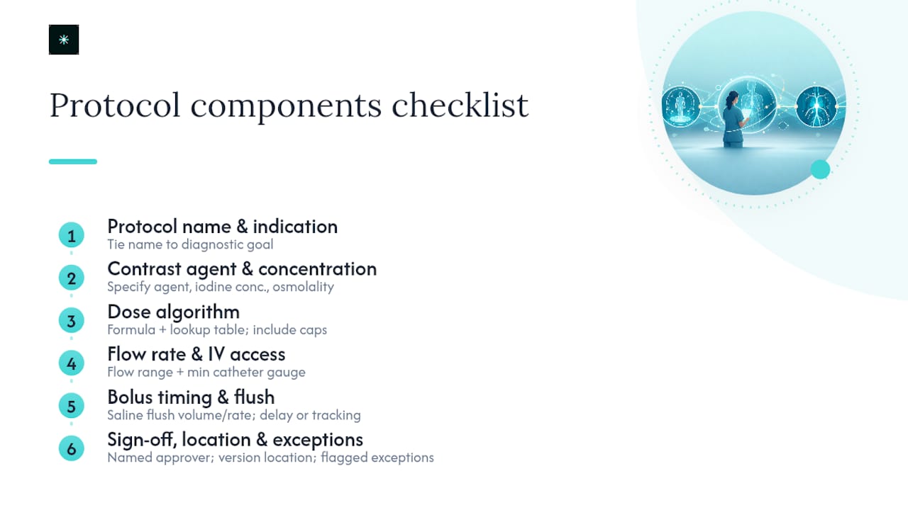
Every protocol document should read like a checklist a technologist can execute without calling anyone for clarification, except in flagged exception cases. That means specificity, not general principles.
Protocol name and indication. Name the protocol after the clinical question it answers, not the body part alone. "CT Abdomen/Pelvis, Suspected Appendicitis" behaves differently than "CT Abdomen/Pelvis, Oncologic Follow-Up," even though both scan the same anatomy. Tie the protocol name to the diagnostic goal so ordering physicians and technologists select the correct one at the point of order entry, not after the patient is already on the table.
Contrast agent and concentration. Specify the exact agent, iodine concentration (for iodinated contrast), and osmolality class your department stocks. Low-osmolar and iso-osmolar agents carry different reaction profiles, and mixing agents across protocols without documenting why creates confusion during pharmacy audits. List contraindications explicitly: prior severe reaction to the same agent class, uncontrolled hyperthyroidism for iodinated contrast, and current nephrogenic systemic fibrosis risk factors for gadolinium-based agents in MRI-adjacent protocols.
Dose algorithm. Most departments choose between weight-based dosing (milliliters per kilogram) and iodine-indexed dosing (grams of iodine per kilogram), with a hard cap regardless of weight. A 250-pound patient does not need double the contrast a 125-pound patient gets; both approaches should include ceiling values, typically somewhere in the range most departments cap around 150 to 200 mL of contrast media for a routine adult CT, adjusted per your radiologist group's own dosing tables. Write the algorithm as a formula plus a lookup table, not a paragraph of prose a technologist has to interpret under time pressure.
Flow rate ranges and IV access. Specify a flow rate range per exam type (for example, 2.5 to 4 mL/s for routine venous-phase studies, higher for CT angiography) along with the minimum catheter gauge that supports it. An 18-gauge or 20-gauge peripheral IV supports higher flow rates than a 22-gauge or 24-gauge line, and the protocol should state what happens when only a smaller gauge is available: drop the flow rate, extend scan delay, or escalate to central access.
Bolus timing and flush technique. Document the saline flush volume and rate that follows the contrast bolus, and note whether your department uses fixed scan delays or bolus-tracking software to trigger acquisition. This detail matters more for CT angiography than for routine venous-phase imaging, but both need a stated default.
Sign-off and location. State who approved the protocol (typically the Imaging Chief or department medical director) and where the current version lives, whether that is a physical protocol book, a PACS-integrated reference, or both. A protocol that exists only in someone's memory is not a standardized protocol.
Pro Tip: Build your protocol templates around the exam's clinical question, not the anatomy alone. Two "CT chest" protocols with different indications often need different contrast timing, and lumping them together is one of the most common sources of avoidable rescans.
How should patients be screened before contrast administration?
Screening should combine a structured questionnaire with targeted lab work, reserving serum creatinine or eGFR testing for patients whose history or risk factors flag them, rather than testing everyone by default. That is the practical reading of ACR guidance and what institutions like UCSF have implemented in their published contrast policies.
Most departments run a two-tier screening model. Tier one is a standardized questionnaire covering prior contrast reactions, known kidney disease, diabetes, metformin use, and pregnancy status for patients of childbearing age. Tier two kicks in when the questionnaire flags a risk factor: that is when eGFR gets pulled, either from a recent lab draw already in the EMR or a point-of-care test if nothing recent exists.
The thresholds matter because they determine action, not just documentation:
- eGFR above 30 mL/min/1.73m²: standard contrast dosing proceeds without special precautions in most protocols.
- eGFR between 30 and 45: many departments require radiologist notification and consideration of reduced dose or alternative imaging.
- eGFR below 30 or active dialysis: contrast use requires case-by-case radiologist review, and non-contrast alternatives get discussed with the ordering physician first.
- Metformin use with impaired renal function: current guidance favors continued use with renal function monitoring rather than automatic discontinuation, but check your institution's own pharmacy-approved pathway since practices still vary.
Pregnancy handling deserves its own line item. For CT specifically, iodinated contrast crosses the placenta, and most protocols require documented pregnancy status before any contrast-enhanced study in a patient of childbearing age, with radiologist consultation for confirmed or suspected pregnancy rather than a blanket refusal.
Premedication regimens for patients with prior allergic-like reactions come in two standard forms, both outlined in the ACR Manual on Contrast Media: a 12-hour oral corticosteroid and antihistamine regimen for elective cases, and a shorter accelerated IV regimen for urgent studies that cannot wait 12 hours. The accelerated regimen carries weaker supporting evidence and does not eliminate reaction risk. It reduces it. Departments running urgent premedication protocols should document that limitation in the consent conversation, not just in the policy binder.
Documentation closes the loop. Scanned or electronically captured screening questionnaires belong in the PACS or EMR record tied to that specific study, not in a separate paper file that nobody can retrieve six months later during an audit. High-risk patients, meaning anyone requiring premedication, dose modification, or radiologist override of a standard contraindication, need documented informed consent that names the specific risk being accepted, not a generic contrast consent form used for every patient regardless of risk tier.
Injection techniques, dosing algorithms, and flow-rate optimization
Dose selection strategies split into three practical approaches, and most departments end up using a hybrid depending on exam type. Fixed-volume dosing (everyone gets 100 mL regardless of weight) is the simplest to implement but produces the widest variability in enhancement quality across body sizes. Weight-based dosing (milliliters per kilogram) corrects for that variability and is the most common approach for routine CT. Iodine-per-kilogram dosing, which calculates the actual grams of iodine delivered relative to patient weight, gives the tightest control over enhancement consistency but requires more setup work in the injector software and works best for CT angiography and other studies where precise vascular timing drives the diagnosis.
Flow rate and catheter gauge need to move together, not independently:
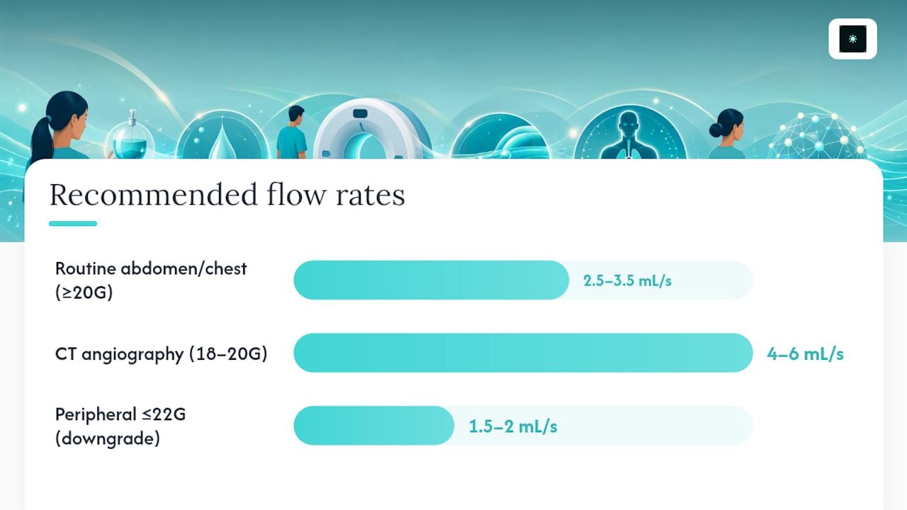
- Routine abdomen/pelvis or chest CT: 2.5 to 3.5 mL/s typically supports adequate enhancement through a 20-gauge or larger peripheral IV.
- CT angiography (chest, abdomen, or extremity runoff): 4 to 6 mL/s is common, generally requiring an 18-gauge or 20-gauge line to sustain that rate without infiltration risk.
- Peripheral access limited to 22-gauge or smaller: down-rate flow to roughly 1.5 to 2 mL/s and extend the scan delay accordingly; document this as a defined exception path rather than leaving technologists to improvise on the spot.
Power injector setup should include a mandatory test flush before the contrast bolus, confirming the line accepts flow without resistance or signs of infiltration. Every injector needs its abort switch clearly accessible to the technologist monitoring the injection, and staff should be trained to stop the injection at the first sign of extravasation: localized swelling, patient-reported burning, or resistance on the injector readout. Extravasation prevention is largely about access site selection (avoiding small hand veins for high flow rates) and real-time monitoring during the first several seconds of injection, when most extravasation events start.
Hand injection remains a valid alternative for pediatric studies, patients with fragile or difficult venous access, and some central-line administrations where power injector use carries higher risk. Central-line contrast administration needs its own written precautions, since not every central line is rated for power injection, and pushing contrast through an unrated line at injector pressures can rupture the catheter.
Pro Tip: Keep a documented "downgrade path" for every high-flow protocol. When a technologist only gets a 22-gauge line on a CTA order, the protocol should already tell them exactly what flow rate and scan delay to use instead of forcing an ad hoc decision mid-exam.
Contrast reaction preparedness and management
Reactions get graded by severity, and the response has to match the grade without hesitation, since the window for effective intervention in severe cases is short.
- Mild reactions (limited hives, itching, or nausea) generally need observation and, if symptoms progress, an oral antihistamine. No interruption of the imaging schedule is usually required beyond the observation period.
- Moderate reactions (diffuse urticaria, facial edema, mild bronchospasm) require immediate physician or designee notification, IV antihistamine, and close monitoring for progression to severe symptoms.
- Severe reactions (laryngeal edema, significant bronchospasm, hypotension, or loss of consciousness) require immediate activation of the emergency response protocol, including epinephrine administration, IV fluids, oxygen, and a call for additional support, following your department's advanced life support pathway.
Every contrast injection room needs a stocked reaction kit within immediate reach, and departments should audit that kit on a fixed schedule rather than assuming it stays complete.
| Reaction kit component | Minimum requirement |
|---|---|
| Epinephrine | Pre-measured doses for both adult and pediatric weight ranges |
| Antihistamines | Injectable and oral formulations on hand |
| Oxygen supply | Immediately available with delivery mask |
| IV fluids | Ready-to-run bags with tubing pre-connected |
| Blood pressure monitor | Functional and calibrated, checked on a fixed schedule |
UCSF's published contrast guidelines require a supervising physician or designee to be available to respond within roughly 5 minutes of a reaction call, a standard that UAB's own contrast policy reflects as well. That response-time expectation should drive staffing decisions directly. If your reading radiologist works from a separate building, or your only qualified responder is off-site, that gap needs a documented backup plan naming a specific alternate responder, not a hope that someone will be reachable.
Staff training has to cover more than a single in-service session. Technologists and nurses working the injection room need periodic reaction-response drills, and the supervising physician group should confirm annually that every listed responder still knows the current kit location and emergency protocol.
Every reaction, regardless of grade, needs post-event documentation: what happened, what was administered, response time from onset to intervention, and outcome. That record feeds directly into the quality assurance review described in the next section, and most institutions also require an incident report routed to risk management for moderate and severe grades.
Governance, protocol approval, and quality assurance processes
Protocol standardization only holds up if someone owns it. Most departments split that ownership three ways: the Imaging Chief or medical director approves clinical content and signs off on changes, the Pharmacy and Therapeutics committee weighs in on agent selection and formulary decisions, and a dedicated QA or Quality Improvement committee reviews performance data and flags protocols that need revision.
The VISN22 contrast policy offers a useful real-world model here: it pairs detailed dosing tables and pre-screening questionnaires with an explicit annual review cycle and assigns responsibility for that review to the Imaging Chief working alongside Pharmacy. That structure keeps the protocol book from becoming a static document nobody revisits until an incident forces the issue.
A working governance cycle includes these fixed elements:
- Protocol book with version control, so every printed or PACS-linked copy shows a revision date and the previous version is archived, not deleted.
- Annual review minimum, with more frequent review triggered by a reaction cluster, a new agent introduction, or a regulatory update.
- Defined approval workflow, meaning a named committee signs off before any protocol change goes live, rather than one technologist's workaround becoming the unofficial new standard.
- KPI tracking built into the review cycle: reaction rates per thousand studies, documented protocol deviations, eGFR documentation completeness, and extravasation rates.
Piloting a change before full rollout catches problems early. Run a revised protocol on one scanner or one shift for two to four weeks, track the same KPIs you use for the standard protocol, and only expand department-wide once the pilot data holds up. Communicate updates through the same channel every time, whether that is a staff meeting, a PACS-embedded alert, or a signed acknowledgment form, so staff are not left discovering a protocol change mid-shift.
Pro Tip: Assign one person, not a committee, to own the protocol book's version history. Committees approve changes; a single named owner keeps the document itself from drifting out of sync with what staff actually use.
Integration with EMR and PACS: workflow enforcement and decision support
Forced fields at order entry catch more errors than any amount of staff training alone. Requiring the ordering clinician to enter indication, allergy history, pregnancy status, and eGFR (or a documented reason it is not available) before the order transmits closes the most common gap: incomplete information arriving at the scanner with the patient already checked in.
Order-entry decision support that ties indication selection to a single unambiguous protocol code reduces last-minute changes at the console, since the technologist is no longer guessing which protocol the ordering physician meant. When that protocol code flows automatically into the scanner acquisition parameters and the injector's dose and flow presets, the manual re-entry step that introduces most human error disappears entirely.
Metadata capture matters as much as the injection itself. Auto-logging the contrast dose administered, agent lot number, and the administering technologist's identifier directly into the PACS record gives you a searchable audit trail without anyone filling out a separate log by hand. That data becomes the raw material for the KPI tracking described in the governance section above.
Embedding safety checklists into PACS and EMR as forced fields is one of the highest-yield operational changes available for reducing variability and preventing missed contraindications, according to guidance UCSF has built directly into its own contrast workflow.
Over-automation is the real risk on the other side of this. A forced field that blocks order submission entirely when a clinician needs to document a genuine clinical exception, say, contrast administration in a borderline eGFR case that the radiologist has already approved by phone, creates workarounds that defeat the whole system. Build an explicit override pathway: a documented reason field plus a required radiologist co-signature for any exception, rather than a hard block with no way through. The goal is forcing completeness, not removing clinical judgment. Departments that get this balance wrong end up training staff to bypass the system entirely, which is worse than having no forced fields at all.
- Require indication, allergy history, pregnancy status, and eGFR (or documented rationale) at order entry.
- Link each indication to one protocol code shared across the EMR, scanner, and injector.
- Auto-capture dose, lot number, and administering staff ID into the PACS record.
- Build a named override pathway with required co-signature, not a dead-end block.
Practical protocol templates and examples
A usable protocol template fits on a single page a technologist can reference in seconds: indication, contrast type and concentration, dose algorithm, flow rate, catheter gauge, scan timing, contraindications, and the documentation checklist. Below are three condensed starting templates for the highest-volume exam types most CT departments run.
| Field | Routine abdomen/pelvis CT | CTA chest/abdomen | Renal CT (stone protocol with contrast phase) |
|---|---|---|---|
| Indication | General diagnostic, oncologic follow-up | Suspected PE, aortic dissection, vascular runoff | Hematuria workup, mass characterization |
| Contrast type | Low-osmolar iodinated | Low-osmolar iodinated, higher concentration | Low-osmolar iodinated |
| Dose algorithm | Weight-based, capped per institutional table | Iodine-per-kg for vascular timing precision | Weight-based, split-bolus option common |
| Flow rate | 2.5 to 3.5 mL/s | 4 to 6 mL/s | 3 to 4 mL/s |
| Catheter gauge | 20-gauge or larger | 18 to 20-gauge | 20-gauge or larger |
| Common exception | Reduced-dose path for borderline eGFR | Down-rate for limited peripheral access | Non-contrast phase substitution if contraindicated |
The routine abdomen/pelvis template covers the largest share of daily volume in most departments and should be the version every new technologist learns first. Its most common exception is the borderline-eGFR patient who needs a reduced dose and radiologist sign-off rather than an automatic hold on the study.
The CTA template demands tighter timing control, which is why iodine-per-kilogram dosing tends to outperform simple weight-based dosing for vascular studies. Its most frequent real-world exception is the patient with only a small-gauge peripheral line available, which forces the flow rate down and the scan delay out, exactly the kind of decision that belongs in a documented downgrade path rather than left to the technologist's judgment under time pressure.
The renal CT template often uses a split-bolus technique, combining excretory and nephrographic phases into a single acquisition to reduce total radiation exposure. Its main exception path is substituting a non-contrast phase entirely when a patient's renal function rules out standard dosing.
Treat these three templates as living documents, not static forms. Link each one to the governance cycle described earlier so that when a KPI review flags a rising deviation rate on, say, the CTA protocol, the fix gets written directly into the template rather than passed along verbally and lost within a month.
Monitoring, audit, and continuous improvement metrics
A protocol without an audit trail is a policy on paper, nothing more. The essential KPIs worth tracking every month are protocol adherence rate (did the technologist follow the documented dose, flow, and access parameters), reaction rate per thousand contrast-enhanced studies, eGFR documentation completeness for flagged patients, and extravasation incident count.
Trattner et al.'s review on CT protocol standardization supports sampling a defined subset of recent studies rather than attempting a full census. A practical audit design pulls 50 to 100 recent contrast-enhanced CT studies each month, checks them against the documented protocol for dose, flow rate, and eGFR documentation, and rolls the findings into a quarterly trend report for the QA committee.
- Sample 50 to 100 studies monthly for dose, flow, and access adherence.
- Track reaction rate per thousand contrast studies as a rolling quarterly figure.
- Run peer review checks in the style of RADPEER on a subset of interpreted studies to catch quality issues tied to protocol variance.
- Present trend data with specific, actionable recommendations, not just raw numbers, at each QA committee meeting.
Trend data only earns its keep when it changes something. A rising deviation rate on one protocol should trigger a targeted retraining session for the specific staff group involved, not a department-wide memo nobody reads closely. When an audit surfaces a pattern (say, extravasation incidents clustering on a particular injector model or a specific shift), the corrective action needs to close the loop visibly: a documented fix, a re-audit date, and a report back to the same committee confirming the fix worked. Skipping that closing step is how the same problem quietly resurfaces a year later.
Standardizing at scale
Rolling out standardized contrast protocols across a multi-site department works best when you resist the urge to fix everything simultaneously. Start with your highest-volume exam type, routine abdomen/pelvis CT is usually the obvious candidate, pilot the revised protocol on one scanner, measure the KPIs for a few weeks, and scale only then. Trying to standardize every protocol on day one guarantees half of them get built poorly under time pressure.
Bring IT into the conversation before you finalize the clinical content, not after. EMR and PACS integration decisions shape what the protocol can actually enforce, and clinical staff rarely know the technical limits of the ordering system until an IT partner points them out. Identify a super-user on each shift, someone respected by peers, not just management, and let them carry questions back and forth during rollout.
Keep the audit process lean. Automate whatever data capture you can pull directly from PACS metadata, and build the feedback loop so staff hear about a deviation within weeks, not at the next annual review. Standardization that takes a year to show results loses staff buy-in long before it proves its value.
How AstraRad supports standardized contrast workflows
Protocol standardization solves half the consistency problem. The other half is getting a fast, accurate, subspecialist read on every study once the scan is done, and that is where a lot of departments quietly fall short even after fixing their contrast protocols. AstraRad provides final signed reports through board certified subspecialists matched to the specific modality, with contractual turnaround commitments of under 4 hours for STAT cases, with 2-hour and 1-hour STAT available on request at a higher multiplier, and 24 to 48 hours for routine studies. For a department that has just tightened its CT contrast protocol, pairing that operational discipline with equally fast, equally consistent reporting closes the loop from acquisition to diagnosis.
Integration is provided directly with existing PACS systems, so there is no separate portal for technologists or radiologists to manage on top of the workflow changes already covered above.
- STAT CT reads committed under 4 hours by contract, with 2-hour and 1-hour STAT on request.
- Direct PACS integration that fits into the same order-entry and imaging workflow your standardized protocols already run through.
- Per-report pricing structured for departments managing variable volume without long-term overcommitment.
- Peer review and compliance reporting that support the same QA cadence your governance committee already tracks.
If your department is evaluating an outsourced reporting partner as part of a broader standardization push, check AstraRad's coverage and licensing details for your state to see how fast a rollout could realistically move.
This article is general information, not a substitute for advice from a qualified doctor. Consult a qualified healthcare professional about your own circumstances before acting on anything here.
Sources
- Enhancing patient safety: standardization of CT contrast administration (JACR / PubMed)
- CT and X-ray contrast guidelines information (UCSF)
FAQ
What is the ACR Manual on Contrast Media used for?
It is the primary US consensus reference for contrast safety, dosing, and reaction management, and most institutional policies, including UCSF and UAB's, build their own protocols around its recommendations, as detailed in the ACR Manual on Contrast Media.
How fast should a supervising physician respond to a contrast reaction?
Institutional policies from centers like UCSF require a supervising physician or designee to be available to respond within roughly 5 minutes of a reaction call, and staffing plans should be built around that window.
Should every patient get an eGFR test before contrast?
No. Most protocols use a screening questionnaire for all patients and reserve eGFR testing for those flagged with kidney disease, diabetes, metformin use, or other risk factors identified during screening.
How often should contrast protocols be reviewed?
An annual review is the standard minimum, with additional review triggered by a reaction cluster, a new contrast agent, or a change in regulatory guidance from bodies like the ACR.
Can teleradiology support contrast protocol standardization?
Yes. A teleradiology partner like AstraRad integrates with existing PACS systems and delivers subspecialist reads on a committed turnaround, which supports the same consistency and QA goals that standardized contrast protocols are built to achieve.
Recommended
Related on AstraRad
- Resources
2027 Medicare fee schedule: the radiology numbers explained
The CY2027 Medicare fee schedule cuts both conversion factors while ACR estimates radiology gains 2 percent. Both are true. Model your dollars per RVU.
- Resources
Clinician-radiologist communication: closing the loop
Clinician-radiologist communication fails when critical results reach a system but never reach the physician, and closed-loop standards close that gap.
- Resources
The hidden cost of the prelim-plus-overread model
The prelim-plus-overread model pays two radiologists and bills one report per overnight study. Cost the overread time, callbacks, and amendments first.
Put a radiologist's name on your next read.
Tell us your modalities and monthly volume. A complete per-report rate card, with turnaround tiers and SLA terms in writing, lands in your inbox within one business day.