Radiation dose monitoring: a practical program guide
Discover how effective radiation dose monitoring enhances patient safety, optimizes protocols, and ensures reliable dose management in healthcare.

Radiation dose monitoring is a continuous program that captures modality-specific dose metrics, flags outliers against diagnostic reference levels, and drives protocol optimization across a department. The first step is technical: confirm automatic DICOM RDSR capture from every modality, then set DRL-based alert thresholds. From there, a real program rests on three pillars: reliable data capture, governance that assigns accountability, and a personnel dosimetry track that runs in parallel.
TL;DR:
- Accurate documentation of dose metrics like DLP and SSDE is essential for meaningful cumulative dose assessment and clinical decision-making.
- Automated data flow from RDSR and supportive platforms should support size-specific, organ-dose, or Monte Carlo estimates to improve patient safety and protocol optimization.
- Staff radiation exposure monitoring must be integrated into operational workflows with clear ownership, regular review, and correlated with patient dose data for comprehensive safety.
- Protocol review and registry benchmarking are key to reducing patient dose over time, with continuous adjustments based on local data and national comparisons.
- Many dose-monitoring programs fail at follow-up actions, making governance, accountability, and active investigation of alerts critical to effective dose management.
Table of Contents
- Why Radiation Dose Monitoring Matters and What a Program Should Achieve
- What Do CTDIvol, DLP, and Effective Dose Actually Mean?
- How Do CT, Fluoroscopy, X-Ray, and Nuclear Medicine Each Report Dose?
- How Should Facilities Monitor Staff Radiation Exposure?
- What Do Dose-Management Platforms Actually Do?
- How Do You Build and Run a Dose-Monitoring Program?
- How Much Can Dose Optimization Actually Reduce Patient Exposure?
- What Are the Biggest Technical Pitfalls in Dose Monitoring?
- How Does Teleradiology Reporting Fit Into a Dose-Monitoring Workflow?
- Where to Find Authoritative Dose-Monitoring Guidance
- Where Dose Programs Actually Break Down
- Sources
Why Radiation Dose Monitoring Matters and What a Program Should Achieve
Patients who undergo repeated CT, fluoroscopy, or nuclear medicine studies accumulate dose across visits, sometimes across years and multiple facilities. A single chest CT rarely raises concern. A trauma patient who gets three CTs in one admission, or an oncology patient on a surveillance schedule, is a different calculation entirely. That's the practical reason cumulative-dose tracking exists: not to police individual scans, but to give clinicians the full picture before ordering the next one. Tracking individual dose history is changing how justification decisions get made, shifting the question from "how many studies has this patient had" to "how much dose has this patient actually absorbed."
Regulatory pressure reinforces the clinical rationale. OSHA requires employers to monitor radiation exposure when workers are likely to receive specified levels of ionizing radiation, and it directs facilities toward engineering controls, administrative controls, and personal protective equipment as the standard control hierarchy. The Nuclear Regulatory Commission sets its own occupational dose limits for licensees, and the Joint Commission increasingly expects documented evidence that a facility tracks and acts on dose data rather than just collects it. A handful of states have gone further and written CT dose-recording mandates directly into law.
Beyond compliance, a monitoring program earns its keep operationally. Once dose data flows automatically, a physics team can:
- Identify protocols that consistently run above the diagnostic reference level for a given exam type.
- Spot individual technologists or scanners whose parameters drift from the department standard.
- Benchmark performance against national registries instead of guessing at what "reasonable" looks like.
- Build the evidence trail regulators and accreditors ask for during survey.
None of this happens with spreadsheets and manual chart review at scale. It happens when RDSR data flows automatically into a system built to flag it, which is why the technical foundation matters as much as the policy language sitting on top of it.
Our interest here is specific and worth stating plainly: AstraRad reads studies and does not sell dose-management software. A monitoring program produces the numbers, and the interpretation workflow around it decides whether anyone acts on them. AstraRad routes every study by modality and body part to a fellowship-trained subspecialist from a panel spanning ten subspecialties, which is what lets a dose outlier on a repeat CT reach a radiologist who reads that exam every shift and can say whether the protocol or the indication is the problem.
What Do CTDIvol, DLP, and Effective Dose Actually Mean?
Four terms get used loosely in clinical conversation, but they measure different things, and mixing them up leads to bad comparisons. Exposure measures ionization in air and is largely a legacy term at this point. Absorbed dose (measured in grays) quantifies energy deposited per unit mass of tissue. Equivalent dose (in sieverts) adjusts absorbed dose for the type of radiation. Effective dose takes equivalent dose a step further and weights it by tissue sensitivity, producing a single whole-body risk estimate that lets you compare, say, a chest X-ray to a CT scan on roughly the same scale.
Each modality reports its own working metrics, and physicists need to know which one answers which question:
- CTDIvol (mGy) reflects scanner output for a given protocol, useful for comparing settings across machines.
- DLP (mGy·cm) multiplies CTDIvol by scan length, giving a rough proxy for total energy delivered in a study.
- SSDE adjusts DLP for actual patient size, correcting for the fact that CTDIvol is measured on a standard phantom, not your patient.
- KAP or cumulative air kerma (Gy·cm² or mGy) is the fluoroscopy and interventional equivalent, tracking total radiation output over a procedure.
- Administered activity (MBq or mCi) is the nuclear medicine analog, describing the radiotracer dose rather than an external beam.
Pro Tip: Effective dose is a population-level risk metric, not a patient-specific measurement. It's built from reference-phantom data and standardized weighting factors, so quoting it to an individual patient as "your exact dose" overstates the precision. Use organ dose or SSDE when a specific patient's risk conversation calls for more accuracy.
The distinction between exposure tracking and true dose tracking matters more than it sounds. Counting how many studies a patient has had is not the same as recording the dose metrics from each one, and only the latter supports a defensible cumulative-dose assessment. A facility that logs "patient had four CTs this year" without capturing DLP or SSDE for each study has an exposure count, not a dose record, and it can't answer the question a treating physician actually needs answered.
How Do CT, Fluoroscopy, X-Ray, and Nuclear Medicine Each Report Dose?
Every modality speaks its own dose language, and a monitoring system has to translate all of them into something comparable. Here's what to track by modality:
CT. Track CTDIvol and DLP per series, then convert to SSDE for size-adjusted comparisons, especially in pediatric and bariatric populations where a phantom-based CTDIvol badly misrepresents actual patient dose. Multi-series studies (a chest-abdomen-pelvis protocol run as three acquisitions) create a common pitfall: summing DLP across series without checking for overlapping scan ranges or repeated localizer runs inflates the apparent total. Blind spots also show up when a technologist manually overrides a protocol, since that override may not always propagate cleanly into the RDSR.
Fluoroscopy and interventional radiology. Cumulative air kerma and KAP are the primary metrics, and both need real-time tracking during long procedures, not just an end-of-case summary. Peak skin dose is the metric that actually correlates with injury risk, and it's notoriously hard to estimate from KAP alone because it depends on beam angle, table height, and patient positioning throughout the case. Some vendor-neutral systems now build 3D skin-dose maps that model dose distribution across the body surface, which turns a single cumulative number into an actual injury-risk zone map, a meaningful upgrade for interventional cardiology and neuro-interventional cases where fluoroscopy time regularly runs long.
General radiography and mammography. Exposure index and deviation index are the digital-radiography analogs, flagging when technique falls outside the expected range for a given body part and detector. Mammography adds average glandular dose as its own dedicated metric, tracked separately because breast tissue dose sensitivity doesn't map cleanly onto other exposure indices.
Nuclear medicine and PET-CT. Administered activity (in MBq or mCi) is the primary metric, but timing matters just as much as the number, since decay between dose calibration and injection changes the effective dose delivered. Combined PET-CT studies require dose accounting from both halves of the exam. The CT portion reports through standard CTDIvol and DLP, while the PET portion reports through administered activity, and a complete dose record needs both tracked and attributed to the same patient encounter rather than filed as two disconnected studies.
How Should Facilities Monitor Staff Radiation Exposure?
Personnel dosimetry runs on a separate track from patient dose monitoring, but the two programs share the same underlying commitment to ALARA. Three dosimeter types dominate clinical practice: thermoluminescent dosimeters (TLDs), optically stimulated luminescence (OSL) badges, and electronic dosimeters that give real-time readouts. TLDs and OSL badges are typically read monthly or quarterly and remain the standard for routine occupational monitoring. Electronic dosimeters see heavier use in interventional suites, where staff want an immediate readout rather than waiting weeks for a lab report.
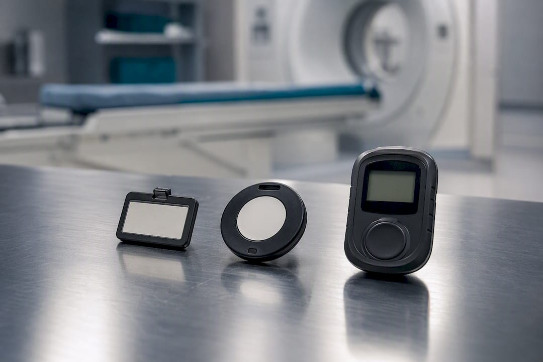
OSHA's guidance on ionizing radiation sets the baseline for employer obligations: monitor workers likely to receive specified exposure levels, and apply engineering controls, administrative controls, and PPE in that order of preference. The NRC sets specific annual occupational dose limits for licensees, and any reading that approaches a meaningful fraction of that limit should trigger a documented investigation, not just a note in a file.
A functioning badge program needs a few operational pieces in place:
- A clear enrollment policy that assigns badges to every staff member with potential occupational exposure, not just the obvious candidates like radiologic technologists.
- A defined cadence for badge exchange and lab turnaround, with a tracking system that flags overdue exchanges before they become a compliance gap.
- Recordkeeping that retains personnel dose history for the duration regulators require, since dosimetry records often need to survive staff turnover and facility ownership changes.
- An escalation protocol that specifies who investigates an elevated reading and what corrective actions get documented.
Pro Tip: Fold personnel dose data into the same QA review cycle as patient dose data. A spike in staff exposure in an interventional suite often correlates with a specific procedure type or a shielding gap that's also worth flagging on the patient side, and reviewing them together catches problems a siloed review would miss.
What Do Dose-Management Platforms Actually Do?
Automated dose-management software exists to solve a scaling problem: no physics team can manually review every study's dose metrics across a department doing tens of thousands of exams a year. These platforms aggregate dose data across every modality and vendor in the department, apply alert thresholds tied to diagnostic reference levels, and generate the dashboards and audit trails that both QA committees and accreditation surveyors want to see.
DICOM RDSR is the backbone. Structured dose reports transmitted in DICOM RDSR format let a facility pull patient-specific cumulative dose history automatically rather than reconstructing it from scanner logs or manual entry. Integration with PACS and RIS means the dose record travels with the study instead of living in a separate silo that nobody checks until an audit forces the question. HL7 feeds from the RIS or HIS add patient demographics and ordering context, which matters when a facility wants to segment dose data by patient age, weight, or clinical indication.
Legacy equipment is the persistent gap. Older fluoroscopy units and some general radiography systems don't emit RDSR at all, so facilities lean on optical character recognition to pull dose values off scanner-generated dose reports or screen captures, backed by manual validation sampling to catch OCR misreads before they corrupt the dataset.
The major platforms in clinical use approach this problem with overlapping but distinct feature sets:
- GE DoseWatch aggregates dose data across multi-vendor fleets, flags studies exceeding DRL thresholds, and supports the reporting workflows accreditation reviews ask for.
- Siemens teamplay Dose builds dose monitoring into a broader fleet-management and analytics platform, useful for health systems already standardized on Siemens imaging infrastructure.
- Sectra DoseTrack operates as a vendor-neutral platform that surfaces dose context directly in the clinical workflow, with configurable DRL and cumulative-dose alerts and support for peak skin-dose modeling in interventional cases.
- Qaelum DOSE focuses on multi-vendor aggregation and alerting with an emphasis on registry-ready reporting for compliance submissions.
Selection criteria should weigh a handful of practical questions: does the platform ingest RDSR natively across your actual equipment fleet, does it support the modality types your department runs heaviest volume on, does it offer size-specific or Monte Carlo-based organ dose estimates rather than relying purely on phantom-based CTDIvol, and does its alerting logic let you set institution-specific thresholds rather than forcing generic defaults. A platform that handles CT beautifully but treats fluoroscopy as an afterthought won't solve an interventional program's actual problem.
How Do You Build and Run a Dose-Monitoring Program?
A dose-monitoring program fails most often not from bad software but from unclear ownership. Governance needs to assign specific responsibilities before the first alert ever fires:
- Medical physicists own protocol review, DRL calibration, and the technical validation of dose data flowing into the system.
- Radiologists weigh in on clinical justification questions, particularly when an outlier alert raises a question about whether a repeat study was warranted.
- Technologists are the front line for catching protocol deviations in real time and flagging equipment behavior that doesn't match expected output.
- Radiation safety officers hold the compliance thread, connecting dose data to regulatory reporting obligations and accreditation documentation.
Once roles are set, a handful of KPIs tell you whether the program is actually working: RDSR coverage rate (what percentage of studies generate a usable structured report), outlier rate against your DRLs, mean time from alert to investigation, and mean dose per protocol tracked over time to catch slow drift before it becomes a pattern.
The operational loop itself is straightforward to describe and harder to run consistently: capture the dose data automatically, flag anything crossing a DRL threshold, investigate the flagged case to determine whether it reflects a clinical necessity or a protocol problem, then either justify the dose or change the protocol, and document the decision either way. That documentation matters as much as the decision itself when a surveyor asks to see evidence of an active program rather than a policy binder that nobody has opened in a year.
Registry participation closes the loop. Submitting data to the ACR Dose Index Registry lets a department compare its protocols against national peer benchmarks rather than relying on published literature that may not reflect current equipment or patient mix, and that comparison is often the fastest way to spot a protocol that's quietly running high.
Pro Tip: Set your local DRLs from your own registry-benchmarked data, not straight from a published national value. National DRLs are a starting reference point, not a target, and a department serving a heavier pediatric or bariatric caseload will need its own adjusted thresholds to avoid chasing false outliers.
How Much Can Dose Optimization Actually Reduce Patient Exposure?
Dose data only earns its value once it drives a protocol change. The concrete levers a physics team can pull include size-based technique factors that scale tube current to patient body habitus instead of running a flat protocol regardless of patient size, iterative reconstruction algorithms that maintain image quality at lower dose, and straightforward protocol consolidation that eliminates redundant or duplicate acquisitions built up over years of ad hoc scanner configuration.
The results compound, though how much depends heavily on how far a department's baseline protocols had drifted from optimized settings before the program started; a department that hasn't reviewed its protocols in a decade has more room to improve than one that reviews annually.
Measuring impact requires a defensible before-and-after comparison, not an anecdotal impression that "things seem better." A cohort comparison across a defined period, matched on exam type and patient size category, gives a physics team a number it can actually defend to a QA committee. Comparing mean CTDIvol or DLP for a given protocol before and after a change, filtered to the same patient size band, isolates the effect of the intervention from natural variation in patient mix.
Benchmarking rounds out the quality-improvement cycle:
- Compare institution-specific mean dose per protocol against ACR Dose Index Registry peer data.
- Set local DRLs at a percentile of your own registry-benchmarked distribution, adjusted for your patient population.
- Re-run the comparison on a recurring schedule, not as a one-time project, since scanner drift and staff turnover erode gains over time.
- Report the trend line to the QA committee in the same cadence as other patient-safety metrics.
Dose management works best treated as a continuous quality-improvement cycle rather than a one-time software installation. A department that installs a platform, sets initial alerts, and never revisits its thresholds is leaving most of the value on the table.
What Are the Biggest Technical Pitfalls in Dose Monitoring?
Data heterogeneity is the problem every physics team eventually runs into. A department running scanners from three manufacturers, spanning a decade of purchase cycles, ends up with wildly inconsistent RDSR completeness and formatting. Data heterogeneity and legacy equipment that doesn't emit RDSR remain among the most persistent implementation obstacles, and the practical fix combines OCR extraction from scanner-generated dose screens with a manual validation sampling process that periodically checks OCR output against the actual image header or a physical measurement.
Normalization strategies help make heterogeneous data comparable. SSDE corrects phantom-based CTDIvol for actual patient size, and Monte Carlo-based calculations improve organ-dose and effective-dose estimates beyond what simple phantom conversion factors provide, particularly for pediatric patients whose body habitus deviates furthest from the standard adult reference phantom used in most conversion tables.
Onboarding a legacy device into a dose-monitoring platform should never happen without validation. A sampling step that checks modality output against ground-truth values, whether the image header, the RDSR itself, or an actual physical measurement, catches systematic ingestion errors before they quietly corrupt months of dose records. Skipping this step is how a facility ends up with a year of dose data it can't trust.
A few other recurring issues deserve a place on any implementation checklist:
- Multi-series CT studies that double-count dose when localizer runs or overlapping scan ranges aren't handled correctly.
- Pediatric patients, where an adult-calibrated DRL badly misrepresents appropriate dose and needs its own size-stratified reference level.
- Effective dose treated as a precise individual risk number when it's actually a population-level estimate built from standardized weighting factors.
- Cumulative dose thresholds confused between a clinical decision point (does this patient need another scan) and a regulatory reporting threshold (does this trigger a compliance obligation), which are different questions with different answers.
Facilities handling patient dose data alongside other protected health information should also treat data governance as part of the technical build, not an afterthought. A HIPAA compliance checklist for systems that collect and transmit dose data is worth reviewing during implementation, since dose records tied to a patient identifier carry the same privacy obligations as any other clinical data.
How Does Teleradiology Reporting Fit Into a Dose-Monitoring Workflow?
Dose monitoring lives inside the imaging department's technical infrastructure, but the reporting workflow surrounding it matters too, and this is where AstraRad's role sits. When a subspecialist reviews a study through a PACS-integrated reporting workflow, the dose history attached to that study through RDSR data can surface at read time, giving the reporting radiologist context on cumulative exposure that's relevant to follow-up recommendations, especially for oncology surveillance or pediatric patients who've had multiple prior studies.
AstraRad's board-certified subspecialists read every study within their trained modality, against a committed STAT tier under 4 hours, with 2-hour and 1-hour STAT on request, and a routine tier of 24 to 48 hours. Studies transfer to the reading radiologist with priors and clinical context attached, and the signed report delivers back into the facility's PACS or RIS over HL7 or FHIR. For dose review specifically, where the workstation sits matters far less than whether the dose record travels with the study: when RDSR data and the priors it belongs to accompany the transfer, the cumulative-exposure picture reaches the radiologist at read time.
It's worth being precise about scope. AstraRad delivers signed interpretations; it does not replace the facility's dose-management platform, its DRL governance process, or its personnel dosimetry program. Those remain the imaging department's responsibility, built on the RDSR capture and QA workflow described throughout this piece. What a reporting partner can do is make sure the dose context a facility has already captured doesn't get lost between the scanner and the final signed report, particularly for combined-modality studies like PET-CT where dose accounting spans two different metrics tracked in two different systems.
Where to Find Authoritative Dose-Monitoring Guidance
A few resources are worth keeping bookmarked for quick reference when a question comes up mid-shift or during a survey prep cycle:
- OSHA's ionizing radiation guidance lays out employer obligations for monitoring occupational exposure and the control hierarchy regulators expect facilities to follow.
- The PMC review on patient dose tracking offers a clear explanation of the distinction between exposure counting and true dose tracking, useful for framing why RDSR-based capture matters.
- The ACR Dose Index Registry gives facilities a peer-benchmarking path for setting institution-specific DRLs rather than relying solely on published national reference values.
- The NRC's occupational dose limit standards set the regulatory ceiling personnel dosimetry programs are built to stay under.
- Vendor product pages for platforms like GE DoseWatch, Siemens teamplay Dose, Sectra DoseTrack, and Qaelum DOSE outline current feature sets for RDSR ingestion, alerting, and skin-dose modeling, worth reviewing directly when evaluating a platform purchase.
Facilities weighing a reporting partner alongside their dose-management build can review AstraRad's teleradiology service coverage or check state licensing details for their specific market before reaching out.
Where Dose Programs Actually Break Down
Most dose-monitoring programs don't fail at the software layer. They fail at the follow-up layer, the part where an alert fires and nobody with the authority to act on it ever sees it in time to matter. A platform can flag every outlier with perfect DICOM RDSR fidelity, and it still won't move a single protocol if the investigation queue sits untouched for six weeks because nobody owns it.
The industry conversation spends a disproportionate amount of energy on which vendor's alerting engine is smarter, and not nearly enough on the unglamorous governance question of who reads the alert on a Tuesday afternoon and actually closes the loop. That's not a software problem. It's an accountability problem, and no dashboard fixes it by itself.
There's also a quieter tension worth naming directly: effective dose gets treated in clinical conversation as a precise, patient-specific number far more often than the underlying methodology supports. It's a population-level risk estimate built from standardized phantoms and weighting factors, and using it to reassure or alarm an individual patient overstates what the number actually claims to know. Organ dose and SSDE get you closer to something defensible for an individual. Effective dose is better reserved for population comparisons and regulatory framing, not bedside conversations pretending to more precision than the math actually provides.
The programs that work best treat the quality-improvement cycle as genuinely continuous, revisiting DRLs against fresh registry benchmarks rather than setting them once during implementation and forgetting they exist. That's a harder discipline to maintain than buying good software, and it's the difference that actually shows up in outcome data.
Sources
Related on AstraRad
- Resources
2027 Medicare fee schedule: the radiology numbers explained
The CY2027 Medicare fee schedule cuts both conversion factors while ACR estimates radiology gains 2 percent. Both are true. Model your dollars per RVU.
- Resources
Clinician-radiologist communication: closing the loop
Clinician-radiologist communication fails when critical results reach a system but never reach the physician, and closed-loop standards close that gap.
- Resources
The hidden cost of the prelim-plus-overread model
The prelim-plus-overread model pays two radiologists and bills one report per overnight study. Cost the overread time, callbacks, and amendments first.
Put a radiologist's name on your next read.
Tell us your modalities and monthly volume. A complete per-report rate card, with turnaround tiers and SLA terms in writing, lands in your inbox within one business day.