Image quality requirements for teleradiology
Image quality requirements for teleradiology: the ACR-based specs, QC checks and contract terms US buyers should set before they send studies.
Image quality requirements for teleradiology come down to one test: a study must reach the interpreting radiologist complete, correctly labeled and at full diagnostic resolution, with no loss of diagnostic information between the scanner and the reading workstation. The ACR,AAPM,SIIM Technical Standard sets the technical rules, and the sending facility's own quality assurance (QA) program has to cover remote exams. This guide turns those rules into a checklist and into contract terms for US hospitals, imaging centers and radiology groups.
- The ACR,AAPM,SIIM Technical Standard allows only DICOM-defined compression (JPEG, JPEG-LS, JPEG-2000 or MPEG) for transmitted and stored images, and it says image data should move at full spatial and contrast resolution.
- Modalities must capture the accession number, patient name and ID, exam date and time, facility name, modality identifier, exam type and orientation at the time of imaging, and DICOM modality worklist is the best-practice way to do it.
- In a 2017 Weill Cornell study, reviewing radiologists agreed with the technologist's reason for rejecting a radiograph in only 64.2% of musculoskeletal cases and 60.9% of chest cases.
- The ACR Practice Parameter for CT and MR at remote locations says facilities should put remote exams inside their ongoing QA, including peer review and periodic audits of image quality, protocol adherence and documentation accuracy.
- A 2021 IAEA remote quality control (QC) methodology paper notes that scanner artifacts can appear suddenly and make an otherwise excellent image useless.
TL;DR
- Diagnostic image quality is a shared job: the sending facility controls acquisition, labeling and transmission, and the radiologist judges whether the study answers the clinical question.
- Write the technical requirements into the contract: DICOM conformance, full-resolution transfer, DICOM-defined compression only, and complete series and prior studies.
- Decide in advance who may request added images or a repeat, and make that a radiologist's call after viewing the whole study.
- Tie the turnaround clock, peer review and QA reporting to the same agreement so image problems never turn into disputes over who owns the delay.
Table of Contents
- What are the image quality requirements for teleradiology?
- Why are we writing about image quality? Our commercial interest
- Who is responsible for image quality: the facility or the teleradiology provider?
- What technical specifications should a study meet before it is sent?
- Which image problems make a remote read unreliable?
- Who decides when to request added images or a repeat study?
- How do display, calibration and compression affect diagnostic quality?
- How should you test image quality before a teleradiology go-live?
- What image quality terms belong in a teleradiology contract and SLA?
- How does peer review support image and report quality?
- FAQ
- Sources
What are the image quality requirements for teleradiology?
Teleradiology image quality requirements are the technical and clinical conditions a study must meet so a remote radiologist can issue a final signed report. They cover acquisition, labeling, compression, transmission, display and the availability of prior studies and clinical history. If any link in that chain degrades the image, the read is at risk.
Diagnostic image quality is the degree to which an image shows the anatomy and pathology needed to answer the clinical question. It is judged by the task, so the same chest film can be adequate for a line check and inadequate for a nodule workup.
The ACR,AAPM,SIIM Technical Standard for Electronic Practice of Medical Imaging applies to any digital image management system, from a single-modality setup to a full PACS to electronic transmission of images between sites for interpretation or consultation. It also says that modality-specific practice parameters and technical standards continue to apply. For a buyer, that means image quality is not a single spec sheet. It is a set of obligations on both sides of the connection.
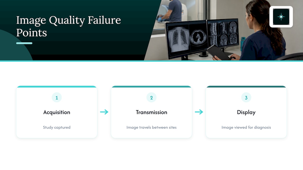
PACS, short for picture archiving and communication system, is the software that stores, routes and displays medical images. DICOM is the international standard format that carries the pixel data and the patient and study information together. Most image quality failures in remote reads trace back to one of three places: how the study was acquired, what happened to it in transit, or how it was displayed.
Why are we writing about image quality? Our commercial interest
AstraRad is a US teleradiology service that delivers final signed reports read by board-certified subspecialists. We sell per-report teleradiology reads to hospitals, imaging centers and radiology groups, so we have a commercial interest in how buyers define image quality. We would rather you set the requirements clearly before a contract starts than discover them in the first week of reads.
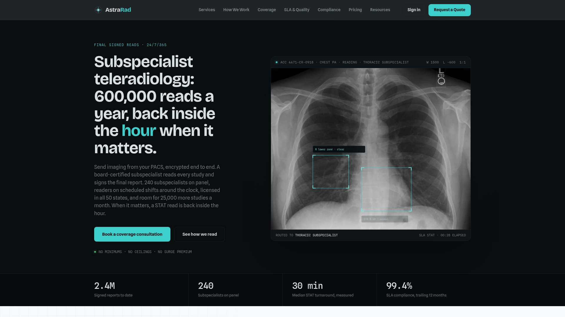
Every study we read is routed to a subspecialist matched to the modality and body part, and reports go straight into your existing PACS with no extra portal, DICOM conformant. Pricing is per report with no minimums. You can see the study types we cover on our teleradiology services page.
Who is responsible for image quality: the facility or the teleradiology provider?
Both parties are responsible, for different parts of the chain. The sending facility owns acquisition, labeling and transmission, and its QA program has to include remote exams. The interpreting radiologist owns the judgment of whether the received study is adequate for the clinical question and says so in the report.
The ACR Practice Parameter for the Supervision and Performance of CT and MR Imaging at Remote Locations states that facilities should include remote examinations in their ongoing QA programs, with peer review and periodic audit of image quality, adherence to protocols and documentation accuracy. In plain terms, outsourcing the read does not outsource the QA program for the images you produce.
That split is why accreditation questions come up in every teleradiology procurement. Our resource on ACR accreditation for teleradiology explains why the facility keeps ownership of QA, and the practical effect is simple: your technologists and physicists still own acquisition quality, while the teleradiology contract should spell out how image problems are reported back to you.
A workable division of duties looks like this:
- Sending facility: scanner and X-ray QC, correct protocols, complete series, accurate patient and exam labeling, DICOM-conformant transmission, and prior studies sent with the current exam.
- Teleradiology provider: subspecialty-matched interpretation, a documented statement of any image limitation in the final signed report, direct communication when a critical structure is not seen, and participation in peer review.
- Both: a feedback channel for image quality, so recurring problems are corrected at the source.
What technical specifications should a study meet before it is sent?
A study should leave your site with complete patient and exam identifiers, full spatial and contrast resolution, DICOM-defined compression only and every series the protocol calls for. These are the controllable items in the ACR,AAPM,SIIM standard, and they belong in the technical exhibit of a teleradiology contract.
The table below converts the standard's language into procurement terms.
| Requirement | What the standard says | What to put in your contract or checklist |
|---|---|---|
| Identifiers | The modality captures accession number, patient name and ID, exam date and time, facility name, modality identifier, exam type and orientation at the time of imaging | A header check on a sample of studies before go-live and after any scanner change |
| Worklist | DICOM modality worklist services are best practice for getting that information electronically | Worklist enabled on each modality that feeds the reading service |
| Resolution | Image data is transferred at full spatial and contrast resolution using DICOM | Full-resolution transfer, with no downsampled copies sent for official reads |
| Compression | Only DICOM-defined algorithms (JPEG, JPEG-LS, JPEG-2000, MPEG); the responsible physician selects and periodically reviews type and amount per modality | A named compression type per modality, reviewed on a set schedule |
| Dose data | Modalities using ionizing radiation should provide a DICOM Radiation Dose Structured Report | Dose reports sent with CT and fluoroscopy studies where supported |
| Change control | Acceptance and integration testing whenever software is introduced or modified, including PACS, RIS, viewers, modalities and reporting systems | A test study run through the full path after every upgrade |
| Monitoring | Documented policies for acquisition, transmission, display and archiving, plus repeat/reject analysis and image quality monitoring | Quarterly QA review that includes remote exams |
All of these rows come from the ACR,AAPM,SIIM Technical Standard. The standard also says that proprietary or nonstandard compression reduces interoperability, and that decompressing and recompressing during data migration causes significant image quality degradation. Our buyer's guide to image compression for teleradiology covers how to set that term in a contract.
DICOM modality worklist is a service that sends scheduled patient and exam details to the scanner so the technologist selects them instead of typing them. It removes the most common source of mislabeled studies.
Which image problems make a remote read unreliable?
The problems that most often stop a reliable remote read are missing series or views, motion, artifacts, wrong positioning, inadequate exposure or contrast timing, and wrong or missing labels. Each one either removes information the radiologist needs or makes the findings ambiguous. In a remote setting nobody can walk to the scanner and ask.
These are the categories to build into your technologist checklist:
- Incomplete coverage: a missing sequence on an MRI, absent reformats on a CT, or a chest film that cuts off the lung apex.
- Motion: blur that hides small lesions or mimics pathology.
- Positioning: a poor lateral view or rotated projection that changes how structures overlap.
- Exposure and contrast: underexposed or saturated radiographs, or contrast phases that miss the target.
- Labeling: wrong laterality, wrong patient, or an orientation marker missing.
- Equipment artifacts: detector or coil faults that affect part or all of the image.
Equipment artifacts deserve special attention because they hide in routine volume. The IAEA remote and automated QC methodology notes that artifacts or nonuniformities in the signal can make an otherwise excellent image useless and can occur suddenly. It recommends QC programs that include artifact and uniformity analysis, so local staff can read the image or send data to a remote center for advice, with visual review to make sure artifacts cannot hide pathology.
The practical lesson is that a weekly or monthly QC schedule is a floor. If a radiologist reports a repeating artifact in a series of studies, your contract should route that feedback to the person who can service the scanner.
Who decides when to request added images or a repeat study?
A radiologist should decide, after viewing the questionable image in context with the whole study. Technologist-directed repeats are often unnecessary: a 2017 Weill Cornell study found that for 77.9% of rejected musculoskeletal radiographs and 93.1% of rejected chest radiographs, the clinical question could have been answered without repeating.
The Weill Cornell study of technologist-directed repeat radiographs also found that reviewing radiologists agreed with the reason for rejection in only 64.2% of musculoskeletal and 60.9% of chest cases. Only 28.4% and 3.4% of repeats added helpful information. Unneeded repeats add radiation, patient inconvenience and delay, and in a remote workflow they also stretch the turnaround clock.
When added images are needed, a documented process prevents duplicate or orphaned studies. The University of Alabama at Birmingham's added images policy is a useful model. If the exam is not complete, or the radiologist asks for added images before the final report, the technologist files them under the same accession number. If the patient has already left the table, the technologist must get clear direction from the radiologist, and any repeat-contrast request must be documented with the radiologist, the contrast amount and the specific series.
For a remote relationship, turn that into a short written protocol:
- The radiologist identifies the limitation and states what is missing.
- The radiologist contacts your site through a named channel for time-sensitive cases.
- Your technologist adds images under the original accession number whenever the exam is still open.
- The communication and the decision are documented in the record.
When a study cannot be completed, the final signed report should describe the limitation and recommend the next step. A final signed report is the radiologist's signed, definitive interpretation, as opposed to a preliminary read, and our explainer on STAT, preliminary and final reads covers why the distinction matters for contracts.
How do display, calibration and compression affect diagnostic quality?
An image can be perfect at the scanner and still fail at the reading workstation. Diagnostic monitor performance, calibration and compression all change what the radiologist actually sees, so each needs a defined owner and a schedule in the agreement.
A diagnostic monitor is a high-grade display calibrated for primary interpretation of medical images, as opposed to a standard office screen. The ACR,AAPM,SIIM standard expects documented policies that cover display as well as acquisition, transmission and archiving, and it lists verification of correct diagnostic presentation, meaning image ordering, window/level, sizing and orientation labels, as part of ongoing QA. Window/level is the control that sets which range of image brightness values is shown and how contrast is spread across them.
For teleradiology, ask for these items in writing:
- Where the radiologist reads, and how the display is calibrated and checked.
- Who holds calibration records and how often they are reviewed.
- Whether hanging protocols, orientation and window/level presets display your studies as intended.
- Which compression type is used for each modality, and who approved it.
Our resource on diagnostic monitor calibration goes through the technologist-facing side of that routine. Radiation dose is the other quality dimension that crosses organizations, and our guide to radiation safety policies for teleradiology shows how dose reporting fits the same agreement.
How should you test image quality before a teleradiology go-live?
Run a structured acceptance test before the first live read: send a representative set of studies through the exact production path, check what arrives, and fix every mismatch before volume starts. The ACR,AAPM,SIIM standard requires acceptance and integration testing whenever software is introduced or modified, and it notes that user acceptance testing details can be written into purchase contracts.
A go-live test plan that fits a mid-size imaging center or hospital:
- Choose the sample. Include each modality and the highest-risk study types: multiphase CT, MRI with many sequences, mammography and studies with large series counts.
- Check the header. Confirm accession number, patient name and ID, date and time, facility name, modality and orientation on every sample.
- Confirm completeness. Compare series counts and slice counts at the sending PACS with those at the receiving side, and confirm that prior studies and reports are available.
- Verify full resolution. Confirm that official reads use full-resolution data with the agreed compression type, and that nothing is recompressed on the way.
- Check presentation. Have a radiologist confirm ordering, orientation, window/level and sizing look correct for each modality.
- Document it. Record results, owners and any fixes, and repeat the test after any PACS, RIS, viewer or modality software change.
Our page on how teleradiology works from your PACS to a signed report shows where each of those checks sits in the workflow. In our case, the first signed report arrives within 10 business days of countersignature, and reports return to your existing PACS in DICOM-conformant form.
What image quality terms belong in a teleradiology contract and SLA?
A teleradiology contract should state image-related duties on both sides: your technical delivery requirements, the radiologist's duty to report limitations, the escalation path, the QA reporting you receive and the exact point where the turnaround clock starts. An SLA, or service level agreement, is the part of the contract that sets measurable service commitments such as turnaround.
Without that language, a poor-quality study becomes an argument about who caused the delay. Use the table to check a draft agreement.
| Contract item | What to specify | Why it matters |
|---|---|---|
| Technical exhibit | DICOM conformance, full-resolution transfer, DICOM-defined compression, worklist use | Sets a measurable standard for what "acceptable" means |
| Clock start | The point at which turnaround is measured, such as last-image arrival | Prevents partial transfers from starting the clock early |
| Limitation reporting | How image limitations are described in the final signed report | Gives your clinicians a clear next step |
| Escalation | Named contacts and the channel for critical findings or missing structures | Keeps time-sensitive cases from stalling |
| Added images | Same-accession process and who may request them | Avoids duplicate studies and orphaned images |
| QA reporting | Peer review process and the reports you receive | Satisfies the expectation that remote exams are inside your QA program |
| Change control | Test and sign-off after PACS, RIS or modality changes | Matches the standard's acceptance testing requirement |
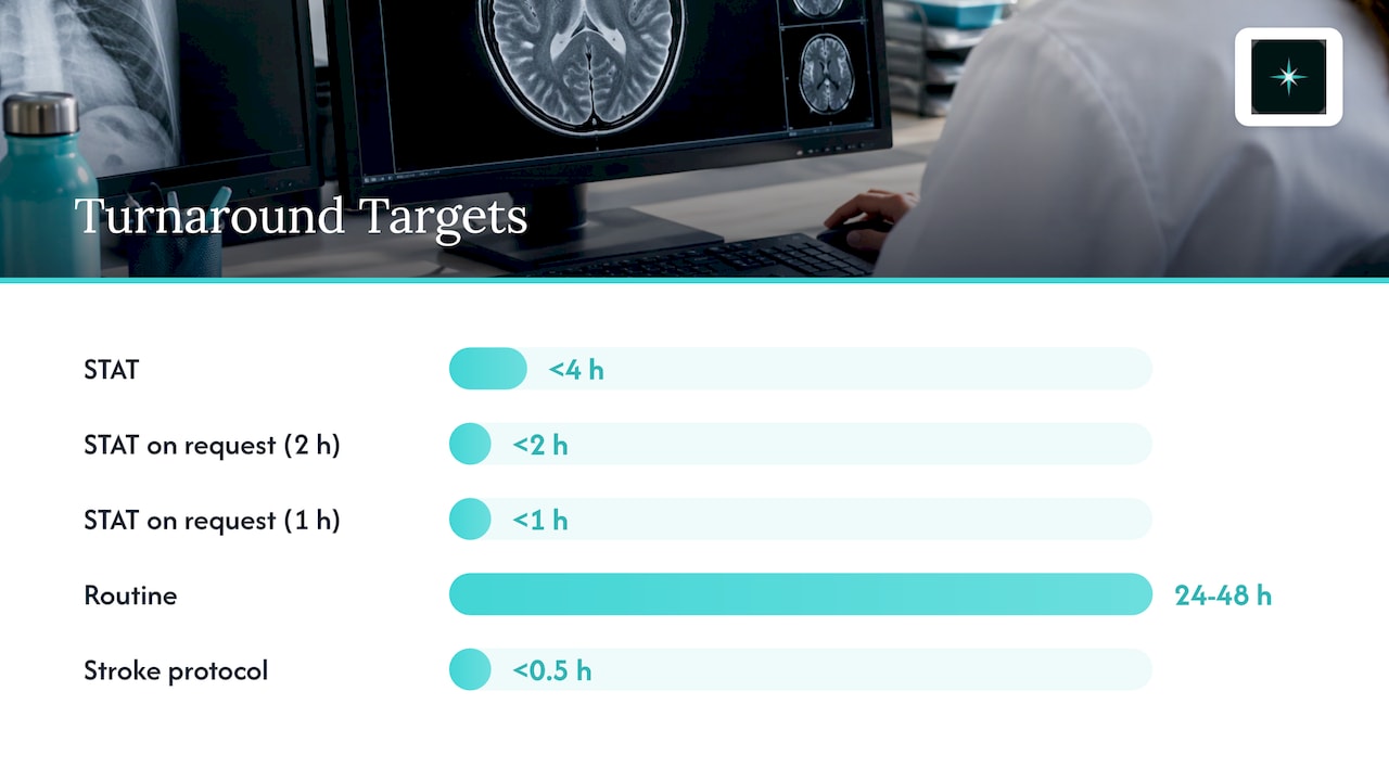
On our side, turnaround is committed by contract and measured from last-image arrival to radiologist signature: STAT under 4 hours (under 2 hours or under 1 hour on request), routine 24 to 48 hours, and stroke protocol under 30 minutes. A clock that starts at last-image arrival matters for image quality, because a study that is still transferring has not yet arrived. For a deeper look at what else to require, see our guide to teleradiologist contracts.
How does peer review support image and report quality?
Peer review is a structured process in which a second radiologist independently re-reads a sample of studies and compares their findings with the original report. It gives you evidence about interpretation quality and a record of whether image problems are being caught.
The ACR,AAPM,SIIM standard lists radiologist peer review among the elements of ongoing QA, alongside repeat/reject analysis, image quality monitoring and turnaround-time monitoring. The practice parameter for remote CT and MR likewise points to peer review within the facility's QA program.
At AstraRad, 1 in 20 reports is independently double-read, blind, for peer review. For your governance committee, the point is to know how a provider's peer review is scored and what you receive from it. Our six-step guide to RADPEER scoring lays out that checklist for radiology teams.
Sources
- ACR,AAPM,SIIM Technical Standard for Electronic Practice of Medical Imaging
- ACR Practice Parameter for the Supervision and Performance of CT and MR Imaging at Remote Locations
- IAEA remote and automated quality control methodology (PMC, 2021)
- Added images policy (University of Alabama at Birmingham, 2021)
Ready to set image and turnaround terms in writing? Book a discovery call or request the per-report rate card.
Frequently asked questions
What is the most important image quality requirement in teleradiology?
The study must reach the radiologist complete and unaltered: correct patient and exam identifiers, every required series, full spatial and contrast resolution, and DICOM-defined compression only. The ACR,AAPM,SIIM Technical Standard frames these as the basics of electronic practice, and a facility's QA program should cover them for remote exams.
Who is responsible for image quality in a teleradiology arrangement?
The sending facility is responsible for acquisition, labeling and transmission, and ACR guidance says it should include remote exams in its QA program. The interpreting radiologist is responsible for deciding whether the study is adequate and for documenting limitations in the final signed report.
How often should we test the image path to a remote reader?
Test before go-live and after any change to a PACS, RIS, viewer, modality or reporting system, because the ACR,AAPM,SIIM standard calls for acceptance and integration testing whenever software is introduced or modified. Add a recurring check to your QA schedule so sudden artifacts and transmission faults are caught early.
Can a technologist decide to repeat an exam on their own?
It is better to have a radiologist decide after reviewing the image in context with the whole study. A 2017 Weill Cornell study found that most rejected chest and musculoskeletal radiographs did not need repeating to answer the clinical question. Put the request process in writing so repeats are intentional and documented.
What should a teleradiology SLA say about image quality?
It should define technical delivery requirements, the point where the turnaround clock starts, how image limitations are reported in the final signed report, the escalation path for missing structures, the added-images process and the QA and peer review reports you receive. Those items keep image problems from becoming turnaround disputes.
Related on AstraRad
- Resources
Turnaround time SLA language for teleradiology contracts
A turnaround time SLA binds only when the contract defines the start event, stop event, statistic, exclusions, and remedy. Clause language for all five.
- Resources
How to audit a teleradiology QA program
Audit a teleradiology QA program with four numbers: double read rate, written discrepancy definition, measured rate, and the workflow after a miss.
- Resources
Nighthawk radiology alternatives: the 2026 shortlist
NightHawk Radiology was acquired by vRad in 2010. Here is what a nighthawk alternative means now, and the overnight coverage shortlist worth calling.
Put a radiologist's name on your next read.
Tell us your modalities and monthly volume. A complete per-report rate card, with turnaround tiers and SLA terms in writing, lands in your inbox within one business day.